
Structure
| Document kind | structure |
| Drawn from | a file — it can also be previewed in the file manager |
| Formats that open here | PDB (.pdb, .ent) · PDBx/mmCIF (.cif, .mmcif) · core CIF (.cif) |
| Refuses to draw below | 260px wide |
What it draws
The molecule in three dimensions — the fold itself, turned with the mouse — beside everything the file states about it: which chains it holds and what kind each is, the sequence of every chain, the ligands and ordered solvent, the disulfides, the residues the depositor declared modified, the unit cell and space group, and the experiment the coordinates came from.
Coordinates are drawn exactly as deposited: the asymmetric unit, in orthogonal ångströms. Nothing here applies the symmetry operators or builds a biological assembly, so the atom count on screen is the atom count in the file.
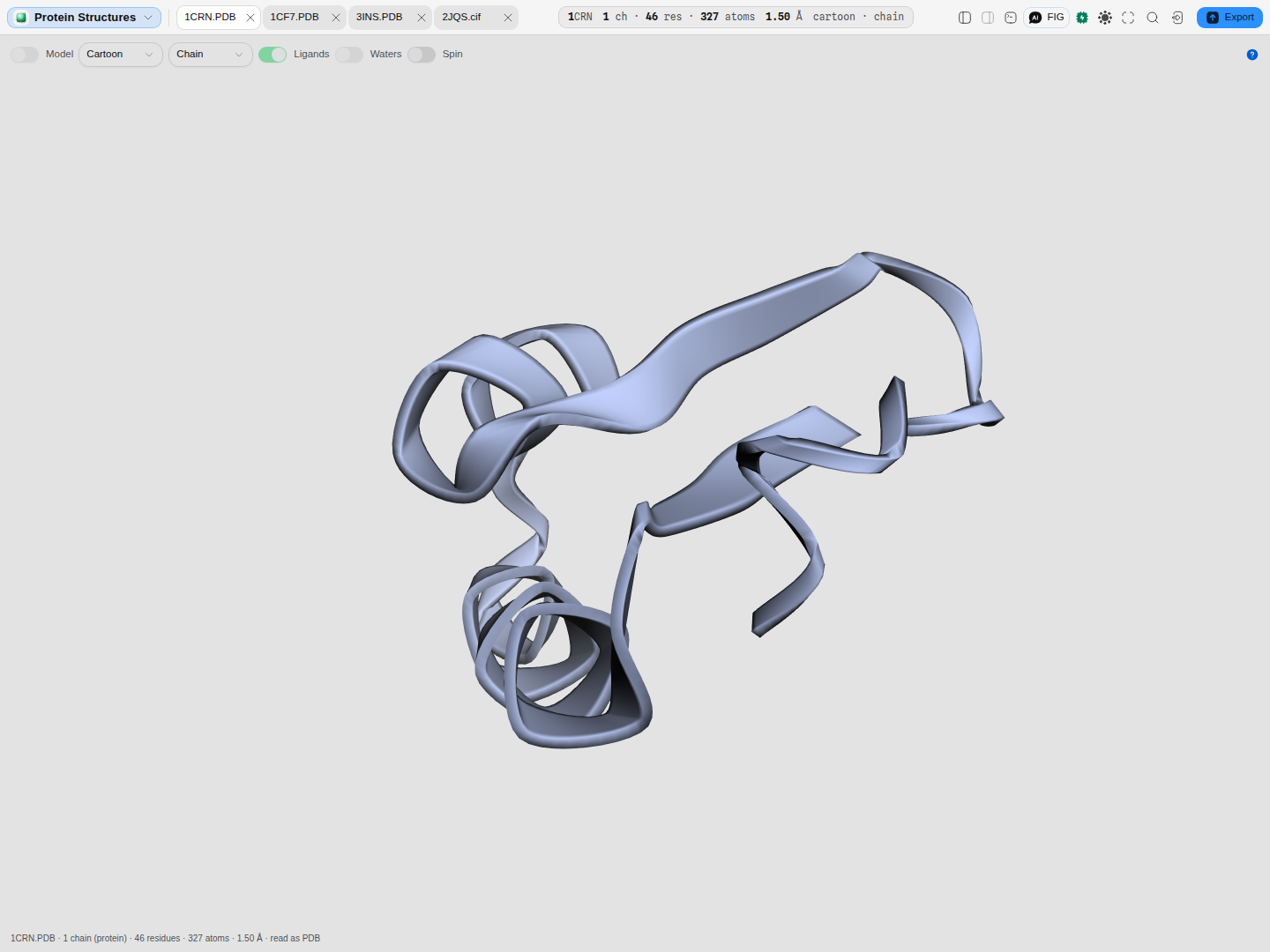 Crambin (1CRN) at 1.5 Å — 46 residues, 327 atoms, one chain. The readings across the top are read
from the coordinate file itself, not from the drawing.
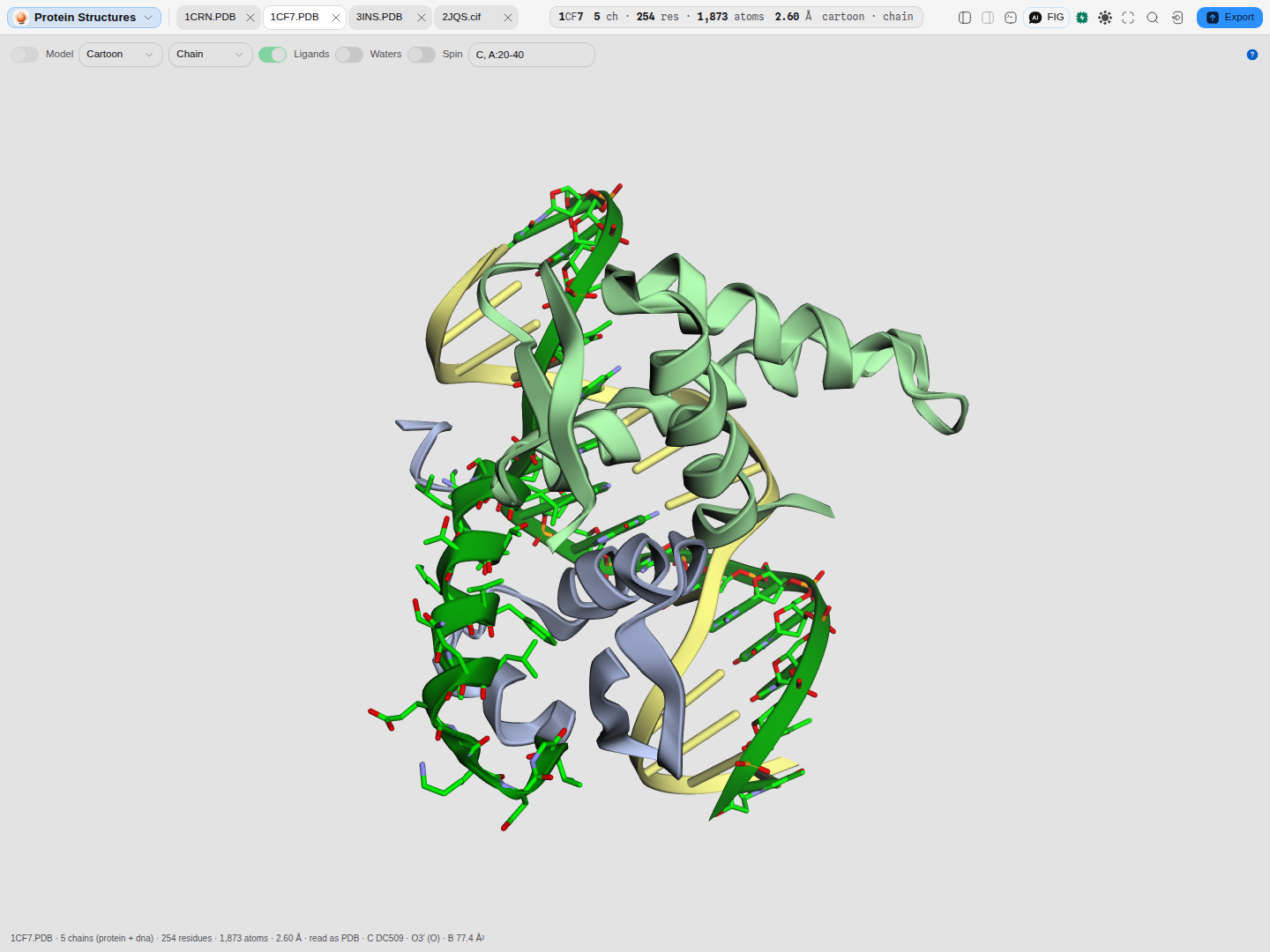
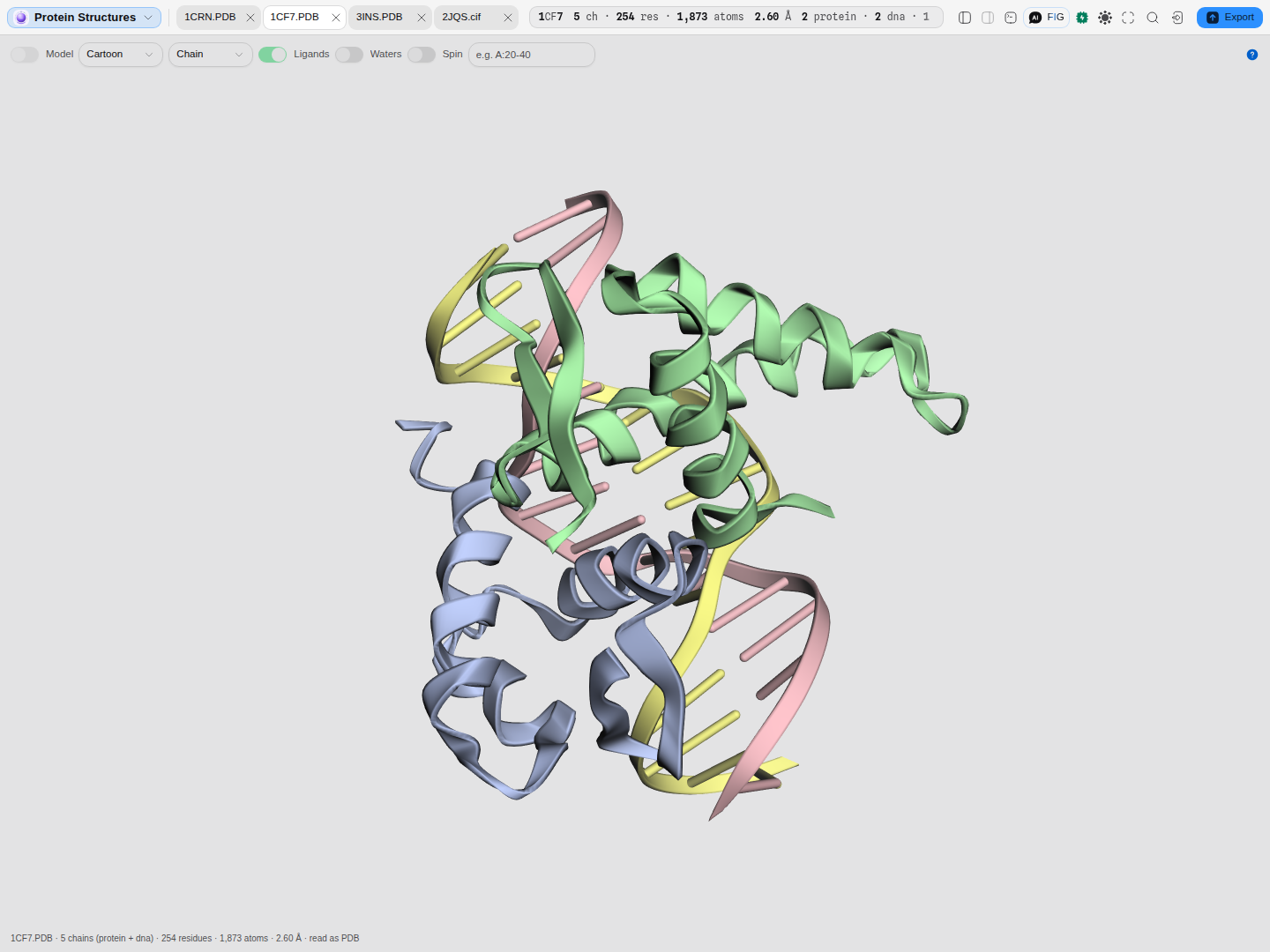 An E2F4/DP2 heterodimer bound to its DNA site (1CF7). Four polymer chains of two kinds: the protein
gets a ribbon, the duplex gets a nucleic-acid cartoon with its base rungs.
What opens in it
Three formats, chosen from the file's contents rather than its name — which matters more here
than anywhere else in the app, because two unrelated dictionaries share the .cif extension:
| Format | What it holds |
|---|---|
PDB (.pdb, .ent) | the fixed-column flat file every older paper cites — coordinates plus the depositor's own annotation |
PDBx/mmCIF (.cif, .mmcif) | the same content as a dictionary, and the only form that can carry an entry too large for the flat file's five-column atom serials |
core CIF (.cif) | a small-molecule crystal structure: a unit cell, a space group, and the symmetry-independent atomic sites in fractional coordinates |
The first two are two serialisations of one archive. They are read the same way and produce the same readings, which is what makes them comparable — and they do not always agree. Where they differ the difference is real: in 1CF7 the older PDB file leaves its 75 waters without a chain identifier while the mmCIF assigns them to chains A–D, so the same 1,873 atoms are five chains in one file and four in the other.
The third is a different dictionary that happens to share an extension. A core CIF has no chains, no residues and no polymer at all, and its coordinates are positions in the crystal's own basis rather than ångströms — so they are placed in space through the cell's orthogonalization matrix before anything is drawn. Every reading that describes a polymer is absent on such a file rather than answered about a wrapper: it reports its sites, the empirical formula the file states, its cell and its space group.
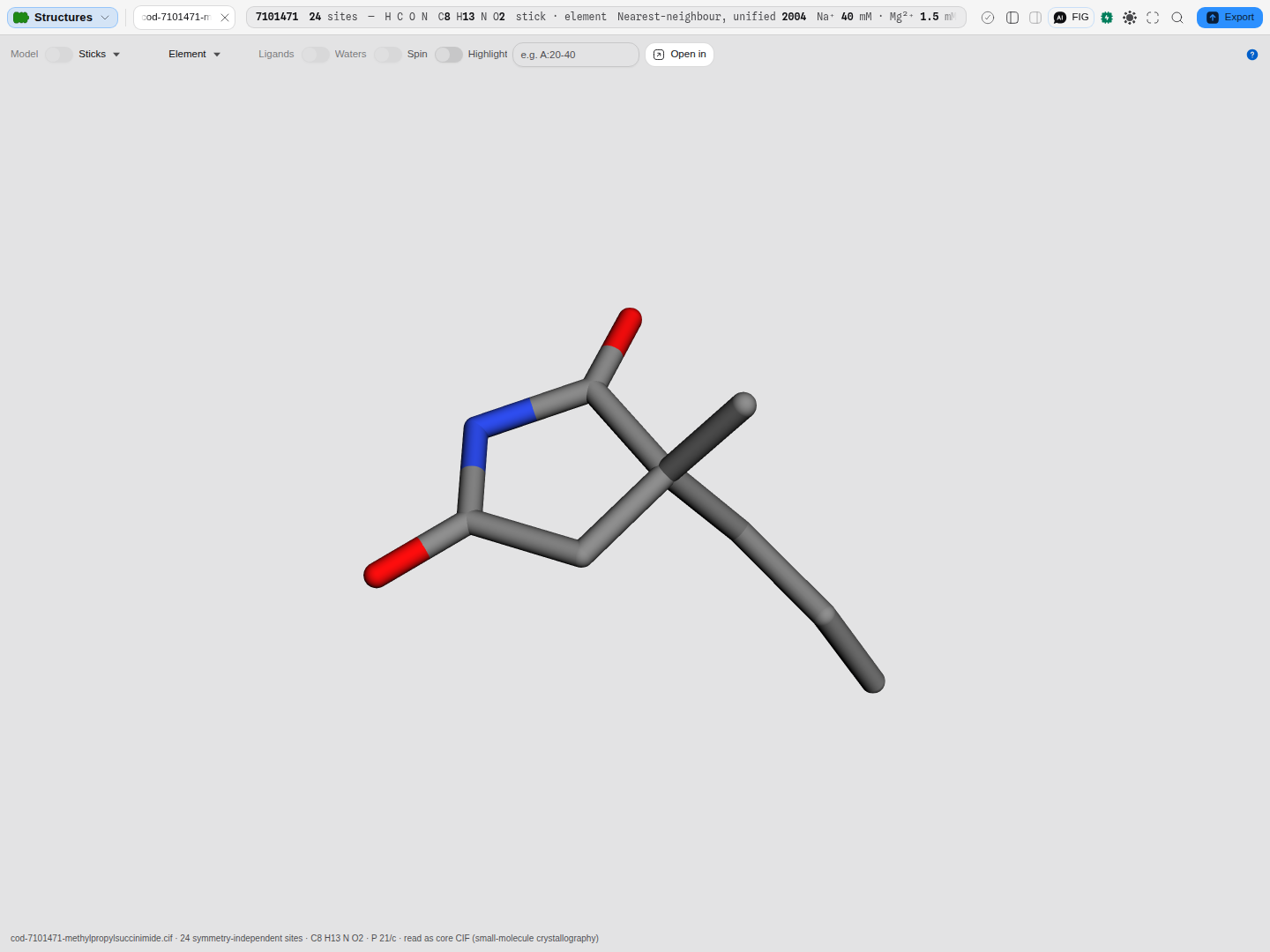 3-methyl-3-propylpyrrolidine-2,5-dione (COD 7101471), monoclinic
P 21/c. Sticks and element
colouring by default, because a cartoon traces a backbone this file does not have — and the reading
says 24 symmetry-independent sites, not "1 chain, 24 residues".
What this computes
Every reading below is derived from the coordinate file on screen, for the model on screen.
- Chain inventory — chains present, the residue and atom count of each, the author numbering range each spans, and its polymer kind (protein, DNA, RNA, water, other). A chain is often mixed, and the label names what most of it is.
- Observed sequence per chain, one letter per residue, beside the construct sequence the file
declares (
SEQRESin PDB,_entity_polyin mmCIF). The two differ wherever a residue was built into the construct but never resolved in the density, which is the gap worth seeing. - Ligand inventory — every non-water heteroatom component, with how many copies of each and how many atoms. Ordered solvent is counted separately and never mixed in.
- Modified residues, with the standard residue each stands in for, taken from the file's own declaration and never guessed from a name.
- Temperature factor (B-factor) range and mean over every atom, in Ų. Absent — not zero — for a determination that states none.
- Alternate conformations: how many atoms carry an alternate-location indicator, which is how you know a side chain is modelled in more than one position.
- Whatever is under the cursor: chain, residue name and number, atom name, element and that atom's own temperature factor.
- Secondary structure: how many helices and strands the file annotates. This is the depositor's annotation, not a calculation over the coordinates, so it says what the entry claims.
- Disulfide bonds declared, with their distance where the file gives one.
- Geometry: the axis-aligned bounding box, its centre, and the longest dimension in ångströms — the number that tells you whether a molecule will fit a pore, a groove or a cryo-EM box.
- Unit cell (a, b, c, α, β, γ) and space group, and the resolution the entry states.
- Elemental composition — how many atoms of each element the model actually holds, counted from the atom sites, most common first. It is the composition question for a structure with no polymer to name chains by, and the only reading that shows hydrogens: a neutron determination places them and an X-ray one at ordinary resolution does not, so their presence is a fact about the experiment.
- Empirical formula, for a file that states one (
_chemical_formula_sum). It describes the contents of the cell, which is why it can name more atoms than the sites the file lists. - Experiment: the technique or techniques, which for a neutron/X-ray structure is genuinely two.
Reading it
- A structure that names itself can be opened where it was deposited. Every reading here is computed from the file in front of you, on your own machine. When the file declares an entry id, the same entry can be opened in the established viewers and entry pages — Mol*, RCSB and PDBe for a PDB or mmCIF entry, the Crystallography Open Database for a small-molecule CIF — seeded on that id. Which is to say: it opens the deposited entry, which can legitimately differ from the file you are holding if that file was edited or stripped. A structure that declares no id has no address to open, and says so; nothing is ever uploaded to make one.
- Representation is a question, not a style. A cartoon answers what the fold is; sticks answer what a side chain, a ligand or an active site is doing; spheres answer how the molecule is packed and what is buried; lines stay legible on a very large model. Ligands and ordered solvent keep their own representation whatever the polymer is set to, so switching a cartoon on never makes a bound zinc invisible.
- Colour answers a different question each way. Chain makes a complex read as its parts. Secondary structure uses the file's own helix and strand annotation. Element is the CPK convention. N to C runs a rainbow along the chain so direction is readable. B-factor runs blue where the model is well ordered to red where the atoms are loosely placed — centred on this structure's mean B, so half the atoms read cooler than typical for this model and half warmer.
- Solvent is off by default, because it surrounds and hides the molecule it belongs to. Turning it on is how you see an ordered water network; leaving it off is how you see the protein.
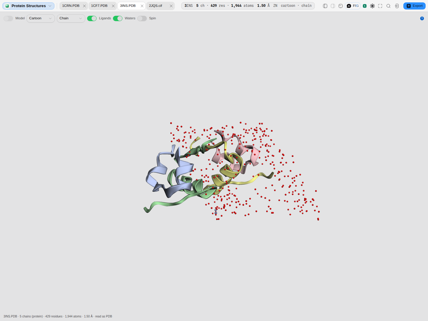 Porcine insulin (3INS) from a neutron/X-ray experiment, ligands and solvent both on. The solvent
here is heavy water, written
DOD rather than HOH — the count is right only because both names are
recognised.
Finding a residue, and asking what one is
Highlight takes the same coordinates the readings beside the picture name, and picks them out of the scene. Terms separate on commas or spaces:
| Term | Selects |
|---|---|
A | chain A, whole |
A:52 | residue 52 of chain A |
A:20-40 | residues 20 to 40 of chain A |
52 · 20-40 | that residue or range in any chain |
ZN · HOH | every residue with that component id |
Numbering is the author's, exactly as the file states it — which is the numbering the paper uses, and may legitimately start below 1 or skip. A selector that reads but matches nothing says so and names what to check; a term that cannot be read is quoted back rather than ignored, because "there is no residue 500 in chain A" and "I could not read what you typed" are different answers.
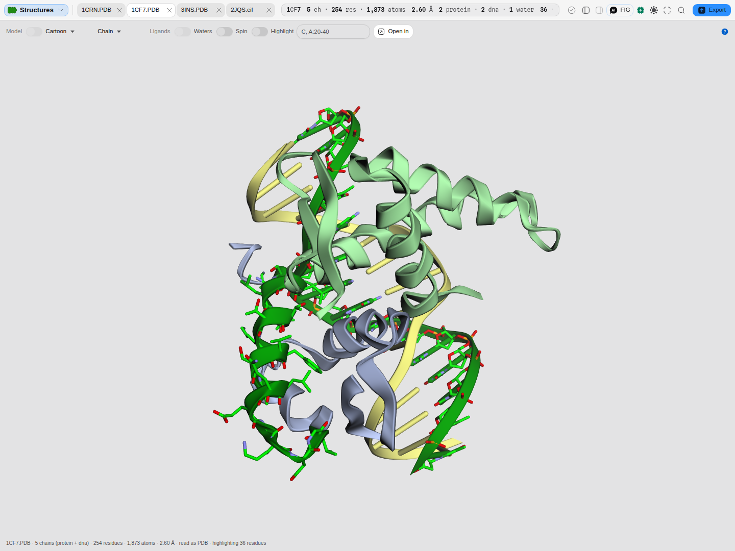
C, A:20-40 on the transcription-factor complex: the DNA strand whole, plus a stretch of the
protein. The count beneath says how many residues were reached.
Hovering answers the question a still picture cannot: what is this. The chain, the residue and its number, the atom name, its element and its temperature factor appear beneath the scene as text — so you can select it, and it never covers what you are looking at.
An ensemble is models, not an average
A solution-NMR entry is deposited as many complete conformers, and they are alternative answers rather than parts of one structure. The Model control draws one at a time or all at once, and the spread between them is the result of the determination. The readings beside the picture always describe the model on screen; they are never summed across an ensemble, because that would multiply every count by the number of conformers.
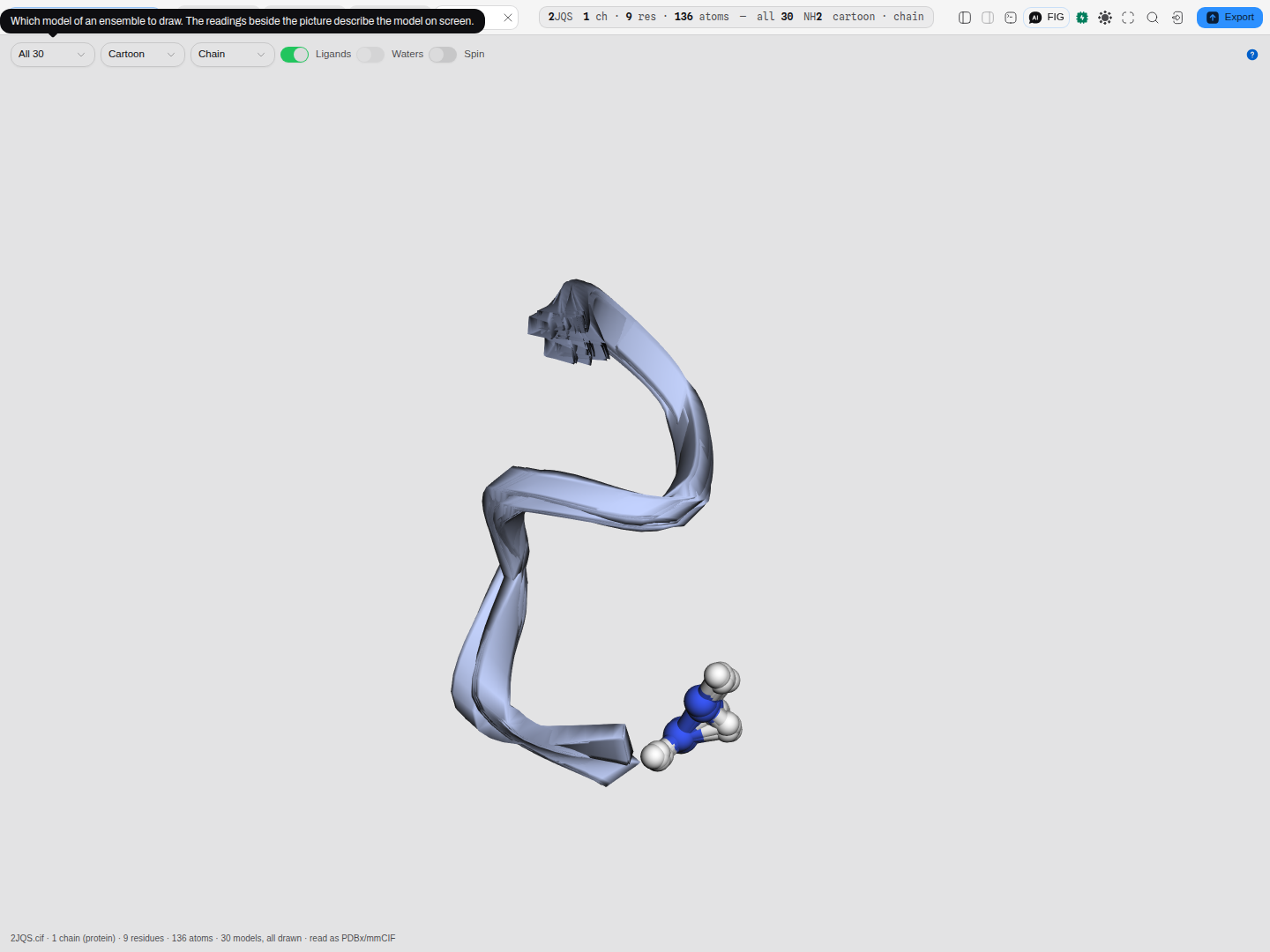 A 30-model NMR ensemble (2JQS) with every conformer drawn.
Such an entry has no resolution, and the reading says so with a dash rather than a number. Colouring by B-factor is offered but not choosable on a file whose atoms all carry zero — the option stays visible and says why, because "this determination has no temperature factors" and "this viewer cannot colour by B" are different statements.
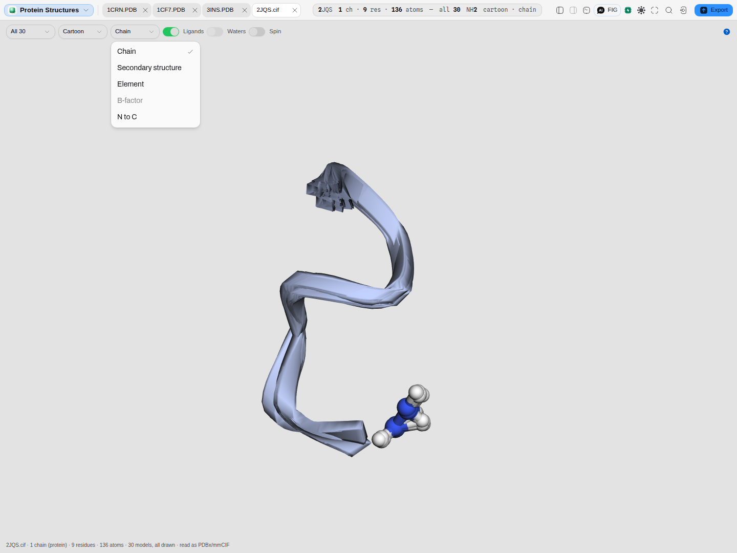 The same ensemble: B-factor present, not choosable, and the resolution reading a dash.
What you can take out of it
Two readings, and they are deliberately a pair.
| What you get | What is in it |
|---|---|
| Chains | One row per chain in the model you are viewing: what it is, how many residues have coordinates, how many the file declares, the difference between those two, the first and last residue number present, and the observed sequence |
| Sequences (FASTA) | The SEQRES the file declares for each chain — the construct that went into the experiment, including residues that were never resolved |
The difference between the two columns in that first table is the disorder, and it is why both are offered. A chain read off the coordinates alone has holes in it wherever the density was too weak to model, and nothing in the sequence you get that way says a residue is missing. In most crystal structures the two numbers disagree.
Coordinates are not rewritten. This canvas reads wwPDB, mmCIF and small-molecule CIF and does not author any of them; the original file is offered back byte for byte instead, which is what a structure is usually wanted for anyway.
Where it appears
As a stage tab, and in the file manager preview. Both use the same viewer, so a structure
reads the same way whichever door you came through. A structure file also offers a second door to its
raw text — a PDB flat file carries a REMARK block, a revision history and the depositor's own
notes that no three-dimensional scene can show.
Performance and limits
- The viewer is loaded on the first structure file you open, never at startup.
- Files over 60 MB are refused rather than loaded, with the size stated. A structure that large is normally distributed as mmCIF; a single asymmetric unit, or one model of an ensemble, will open.
- A file that carries no readable atom records is refused with that reason, naming the first problem found, rather than drawing an empty scene.
- A line the reader cannot parse becomes a stated warning and the rest of the file is still read: one truncated atom record does not make a structure unopenable.
Provenance of correctness
The readers are checked against gemmi, the macromolecular library CCP4 and the Protein Data Bank's own tooling are built on. Every atom of every bundled structure — 11,488 of them across eight files and both serialisations — is compared serial by serial, name by name, and coordinate by coordinate against what gemmi reads from the same bytes, with occupancies and temperature factors at the precision the format carries.
Residue classification is checked against the wwPDB Chemical Component Dictionary, the archive's own register of what every three-character residue name means, so water, amino acids and nucleotides are separated the way the archive separates them rather than by a list assembled here.
The column definitions come from the wwPDB PDB File Format v3.3 specification and the PDBx/mmCIF dictionary, and the extended atom numbering from the hybrid-36 reference implementation. The bundled structures are current or verified revisions from the RCSB Protein Data Bank, each checked against its own deposited revision history.
For a small-molecule crystal structure the arithmetic that places a fractional coordinate in space is the one step that cannot be checked by eye, so it is checked against gemmi element by element and site by site, in all three cell regimes a structure can have: every angle 90°, one angle off 90°, and all three off. The convention itself — which crystal axis is aligned with which Cartesian one — is the one International Tables for Crystallography volume B states for the Protein Data Bank. The bundled examples come from the Crystallography Open Database, each with its own COD entry number.
Related
A structure in a project is usually the protein the rest of the project expresses. The bundled
example is exactly that shape: a small high-resolution protein, a transcription factor caught on the
DNA site it recognises, a hormone with its metal, and an ensemble that shows what a solution
structure actually knows — then three crystal structures that share the .cif extension with none of
its meaning.
A molecule that is not a crystal structure — an SDF, a molfile, a SMILES string, a reaction — is a different canvas with a different question behind it, and opens as a molecule.