
Digest result
| Document kind | digest |
| Drawn from | a tool result — there is no file behind it |
What it draws
The digest result is the answer to what did my enzymes actually do to this molecule, in the two forms that answer is useful in.
The gel, which is what you would see. One lane per enzyme combination you set up, drawn against a marker ladder on the shared agarose physics, so the picture on screen is the picture you are about to compare your real gel photograph against. Everything the gel canvas does is available here: agarose percentage, mobility law, DNA form, custom ladders, your own Rf calibration, and the three render modes.
The fragment table, which is what you would need to act. One row per fragment, sortable, with eight columns: the lane it came from, its Start and End on the parent molecule, its Length, and then the four that decide whether it can be cloned — Left Cutter, L. Overhang, Right Cutter, R. Overhang. The overhang is the half of that answer most tools leave out, and it is the half that decides what a fragment will ligate to.
A digest of a circular plasmid with three enzymes is one lane, six rows and one number you can act on; a digest with a zero-cutter in a second lane is a lane that draws empty and says why.
What opens in it
Nothing opens in it. There is no digest file: the result is drawn from a run of the Restriction Digest tool on the open molecule, and it lasts as long as that result does. To keep it, export it: the fragment table leaves as CSV, and Open in new tab persists that same CSV as a real file in the workspace.
What is not here: the fragments as sequences you can edit. Clicking a fragment selects its span on the molecule it was cut from, where you can inspect it base by base — the result is about the reaction, the map is about the DNA.
What this computes
The digest result computes the reaction, then computes the picture of it.
- Cut positions for every enzyme in every lane, over the parent molecule, respecting whether it is circular or linear — a circular molecule cut once yields one linear fragment, not two.
- The fragment set per lane: each fragment's Start and End on the parent, 1-based and
inclusive of both ends, so
Length = End − Start + 1. The panel states that convention under the table, because1,999 → 2,055 = 57reads as an off-by-one to anyone assuming half-open intervals. - Fragment length in base pairs, per fragment.
- The cutter at each of the two ends —
Left CutterandRight Cutter— so a fragment's provenance is not inferred from its position in a list. - The single-stranded overhang at each end, written as the strand it protrudes on, with blunt reported as blunt rather than as an empty cell. This is the half of the answer that decides what a fragment will ligate to, and it is the half most virtual digests leave out.
- Zero-cutter detection: an enzyme set with no site on this molecule produces a lane that draws empty and is labelled with that reason. An empty lane is never how you discover an enzyme does not cut here.
- Partial-digest fragments, off unless asked for: the additional fragments an incomplete reaction would leave. The panel states how many that adds before you turn it on, because the set is much larger than the complete digest.
- Reaction conditions per lane, because a lane is one tube: the recommended NEBuffer, scored on the worst enzyme's percent activity in it, with any buffer where an enzyme shows star activity ruled out whatever its percent; the activity in every buffer, so the runner-up is there when the digest has to be split; the incubation temperature; a required cofactor (SAM or ATP); and the dedicated buffer some enzymes ship in. When no buffer or no single temperature serves every enzyme in the lane, the reading says so and names each buffer's reason. The activities, star flags and temperatures are New England Biolabs' published figures; the cofactors come from REBASE.
- Heat inactivation, per enzyme, as NEB lists it: 65 °C or 80 °C for 20 minutes, or not heat-inactivatable, in which case the DNA needs a column or gel cleanup before the next enzymatic step. A lane is only heat-killable when every enzyme in it is; the steps are listed per enzyme, never merged into one temperature.
- Everything the gel canvas computes, on the same agarose physics — relative mobility per fragment, topology form factors, ladder calibration, the resolving window and the brightness normalisation — because the plate here is that canvas, not a picture of one.
Reading it
Positions are 1-based and inclusive of both ends. Length = End − Start + 1. The panel states
this under the table rather than leaving you to test it, because 1,999 → 2,055 = 57 reads as an
off-by-one to anyone assuming half-open intervals, and it is not one.
An overhang is written as the strand it protrudes on. Left Cutter plus L. Overhang together
are the claim: this end was made by this enzyme and presents this single-stranded sequence. An
empty overhang cell with blunt beside it is a blunt end, which is a real answer and not a missing
one.
A fragment appearing at a size you did not expect is worth checking against the resolving window before checking against your enzymes. The gel says what the chosen agarose resolves; a fragment outside that window is placed by extrapolation, and the table, not the gel, is where its size is authoritative.
An empty lane means this enzyme set does not cut this molecule. It is drawn empty and labelled with the reason. An empty lane must never be how you discover an enzyme has no site here, so it never is.
Partial digests are a separate question, and they are off unless you asked. With partials on, the lane also carries the fragments an incomplete reaction would leave, which is a much larger set — the panel says how large before you turn it on.
Controls
The result carries its render mode (Bands · Raster · Both), its brightness reference (Whole plate · Per lane) and its Send to Gel Bench action; everything that changes what the result contains belongs to the digest run itself — the enzymes per lane, the ladder, the agarose percentage, the mobility law, the DNA form and the calibration.
Both of those are ways of looking at a saved plate rather than parts of the run, which is why they are offered on a snapshot at all. The brightness reference is never disabled the way Raster and Both can be: re-scaling a plate needs only the two reference values the result already carries.
A saved result tab is a snapshot, and it is honest about what a snapshot can do. A tab saved from a bands-only run carries no raster image, so Raster and Both are offered disabled with that reason rather than hidden: the capability exists, this particular snapshot just cannot show it.
Selection flows both ways, and it lands everywhere at once. Click a band on the gel or a row in the table and that fragment's span is selected on the circular map, on the linear map, in the sequence, in the panel's own target chip and in the status bar. That is one selection, not five copies of one.
Where it appears
In the Restriction Digest panel in the right rail, and as its own stage tab via Open in new tab when you want the fragments beside the map rather than under it. The tab renders the same gel and the same table the panel does, from the same run.
Performance and limits
The fragment table is virtualized, so a digest with a frequent cutter (an AluI on a plasmid is
routinely dozens of fragments) scrolls rather than stalls. The gel draws one image per lane rather
than one element per band.
The limits worth knowing are scientific. Cut positions come from REBASE, and REBASE does not state a cleavage position for every enzyme it lists: an enzyme whose cut is undetermined is shown as (cut site unknown) and is not given an invented blunt cut. Where that happens the tool answers the next question instead, naming the enzymes on the same recognition sequence whose cut position is known.
Screenshots
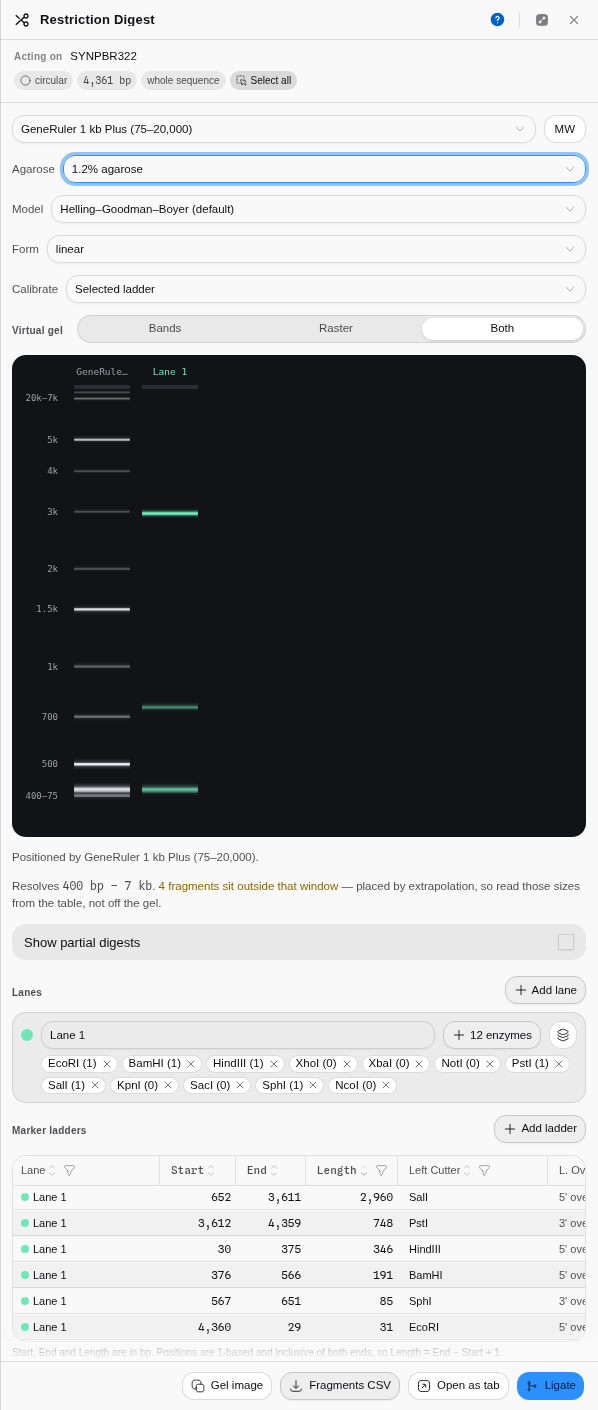
Twelve common cutters loaded into one lane on real pBR322, each chip carrying its live cut count on this molecule, run at 1.2 % agarose in Both mode. The six real fragments are in the table: 2,960 · 748 · 346 · 191 · 85 · 31 bp, and the resolving-window warning is firing because the 31 bp fragment is below what this gel resolves.
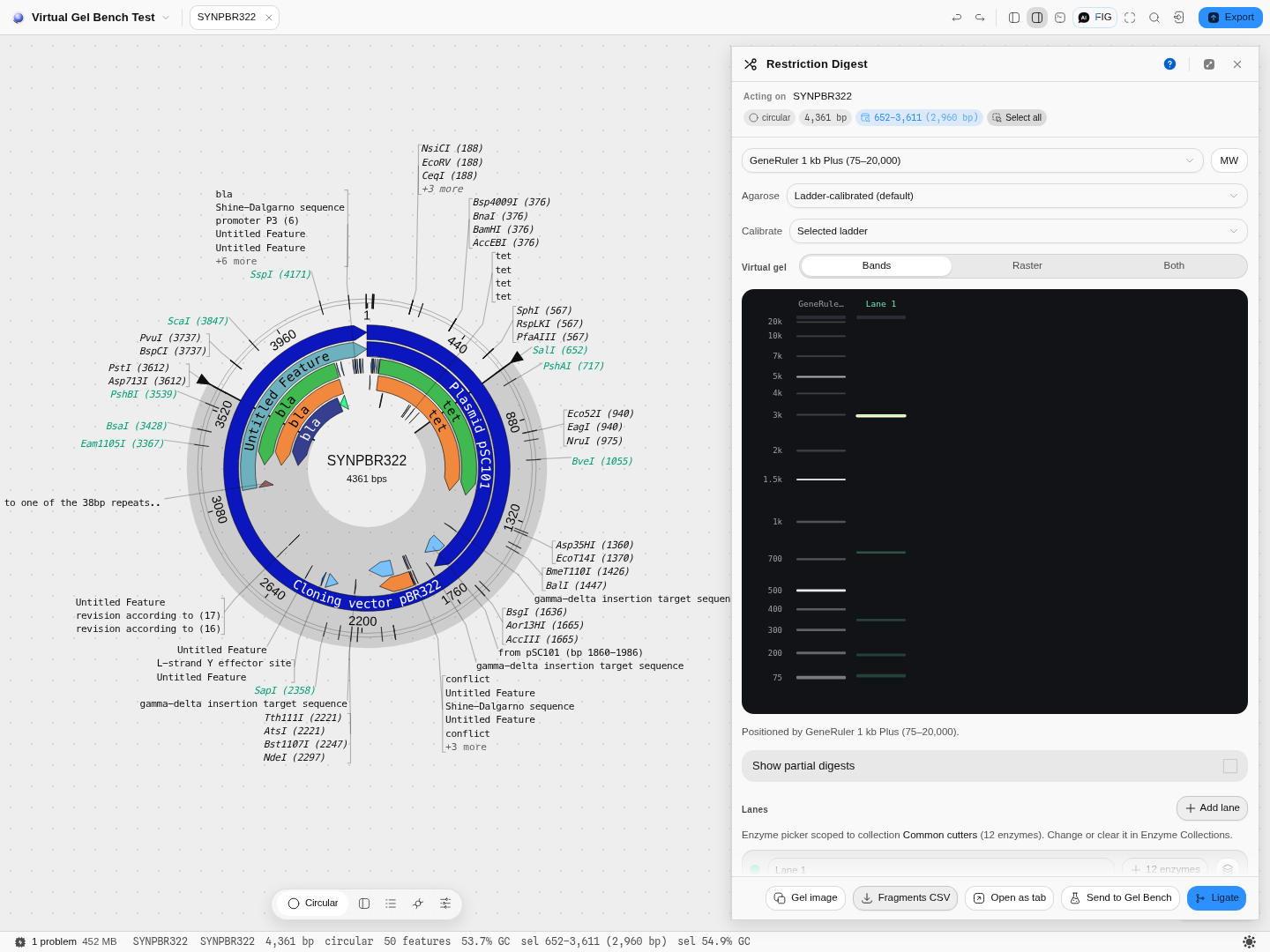
One selection in four places: clicking the 2,960 bp SalI–PstI row highlights its band on the gel,
its arc on the circular map, its chip in the panel, and its span in the status bar as
652–3,611 (2,960 bp).
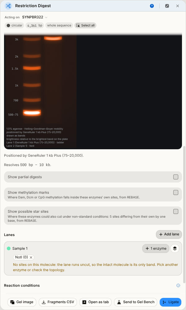
The designed empty state: NotI has no site in pBR322, so the lane is empty and the panel says
that is why.
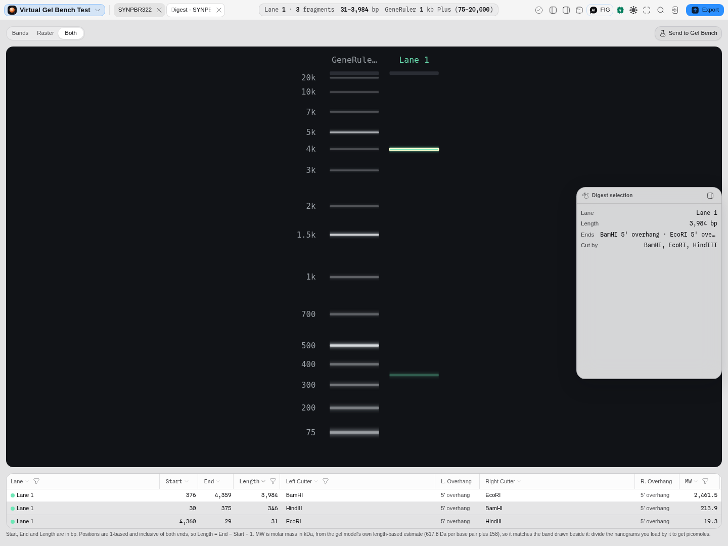
The same digest kept as a tab of its own: pBR322 cut with BamHI + EcoRI + HindIII, gel filling the stage above its fragment table. The tab reopens in the render mode the run was made in — this one was made in Both, so the rectangles are drawn over the raster. The 31 bp fragment is in the table and not on the plate because it runs off the bottom of a GeneRuler 1 kb Plus; the gel does not invent a position for a band its ladder cannot place.
How this is checked
The fragments and their ends come from a digest implementation validated against the published tools, and the recognition sequences and cut positions are generated from REBASE, the authoritative restriction-enzyme database, rather than from a list maintained here. The band placement is the same pydna-validated migration model the gel canvas uses. Where REBASE states no cleavage position, none is shown.
Related
Written by Restriction Digest. Its lanes hand over to the Gel Bench and draw on the gel canvas; the molecule the fragments were cut from is the sequence map. The enzymes themselves are described in restriction enzymes and managed in enzyme collections.
Compare to reference
The multi-sample variant workspace: every position where a sample differs from the reference, beside the aligned bases themselves.
Gel
A gel canvas is an agarose run you did not have to pour: lanes of DNA placed against a molecular-weight ladder by a real, published migration model rather than by drawing bands where they look right.